Tuesday
Jan122021
Top 10 health innovations of 2020
 Great to see our recent Cell paper on brain T cells licensing microglia listed as one of the top 10 health innovations of 2020!
tagged  Liston lab, immunology, neuroscience
Becoming a Scientist

Virus Fighter
Build a virus or fight a pandemic!

Maya's Marvellous Medicine
Battle Robots of the Blood

Just for Kids! All about Coronavirus
Learn about our spin-off, Aila Biotech!

Great to see our recent Cell paper on brain T cells licensing microglia listed as one of the top 10 health innovations of 2020!
A team of immunology experts from Belgium and the UK research organisations have come together to apply their pioneering research methods to put individuals’ COVID-19 response under the microscope. Published today in the journal Clinical and Translational Immunology, their research adds to the developing picture of the immune system response and our understanding of the immunological features associated with the development of severe and life-threatening disease following COVID-19. This understanding is crucial to guide the development of effective healthcare and ‘early-warning’ systems to identify and treat those at risk of a severe response.
One of the most puzzling questions about the global COVID-19 pandemic is why individuals show such a diverse response. Some people don’t show any symptoms, termed ‘silent spreaders’, whereas some COVID-19 patients require intensive care support as their immune response becomes extreme. Age and underlying health conditions are known to increase the risk of a severe response but the underlying reasons for the hyperactive immune response seen in some individuals is unexplained, although likely to be due to many factors contributing together.
To investigate the immune system variations that might explain the spectrum of responses, teams of researchers from the VIB Centre for Brain and Disease Research and KU Leuven in Belgium and the Babraham Institute in the UK worked with members of the CONTAGIOUS consortium to compare the immune system response to COVID-19 in patients showing mild-moderate or severe effects, using healthy individuals as a control group.
Professor Adrian Liston, senior group leader at the Babraham Institute in the UK, explained: “One of our main motivations for undertaking this research was to understand the complexities of the immune system response occurring in COVID-19 and identify what the hallmarks of severe illness are. We believe that the open sharing of data is key to beating this challenge and so established this data set to allow others to probe and analyse the data independently.”
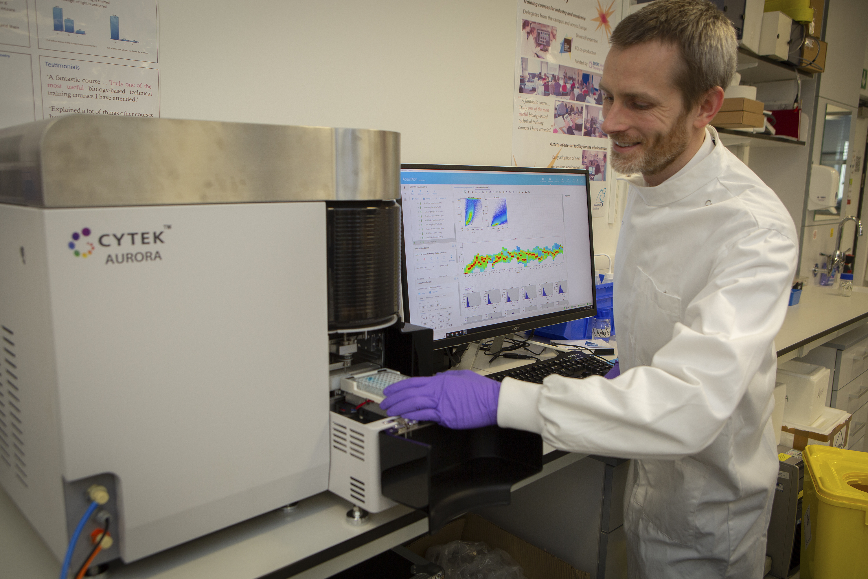
T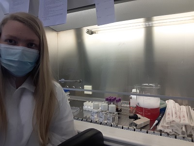 he researchers specifically looked at the presence of T cells – immune cells with a diverse set of functions depending on their sub-type, with ‘cytotoxic’ T cells able to kill virus-infected cells directly, while other ‘helper’ T cell types modulate the action of other immune cells. The researchers used flow cytometry to separate out the cells of interest from the participants’ blood, based on T cell identification markers, cell activation markers and cytokine cell signalling molecules.
Surprisingly, the T cell response in the blood of COVID-19 patients classified as severe showed few differences from the healthy volunteers. This is in contrast to what would usually be seen after a viral infection, such as the ‘flu. However, the researchers identified an increase in T cells producing a suppressor of cell inflammation called interleukin 10 (IL-10). IL-10 production is a hallmark of activated regulatory T cells present in tissues such as the lungs. While rare in healthy individuals, the researchers were able to detect a large increase in the number of these cells in severe COVID-19 patients.
Potentially, monitoring the level of IL-10 could provide a warning light of disease progression, but the researchers state that larger-scale studies are required to confirm these findings.
“We’ve made progress in identifying the differences between a helpful and a harmful immune response in COVID-19 patients. The way forward requires an expanded study, looking at much larger numbers of patients, and also a longitudinal study, following up patients after illness. This work is already underway, and the data will be available within months,” says Professor Stephanie Humblet-Baron, at the KU Leuven in Belgium.
 “This is part of an unprecedented push to understand the immunology of COVID-19”, concludes Professor Liston. “Our understanding of the immunology of this infection has progressed faster than for any other virus in human history – and it is making a real difference in treatment. Clinical strategies, such as switching to dexamethasone, have arisen from a better understanding of the immune pathology of the virus, and survival rates are increasing because of it”.
Professor Liston and Professor Humblet-Baron both emphasized the importance of the scientific team that led the study. "This work happened during a period of incredible stress. When much of our laboratory was shut down due to the pandemic, Dr Teresa Prezzemolo and Silke Janssens were in the hospital day-after-day, preparing blood samples that were critical not just for this study but for a whole host of clinical trials on COVID-19 based in Leuven. Julika Neumann and Dr Mathijs Willemsen put their PhD research on hold to run samples, and Dr Carlos Roca and Dr Oliver Burton provided the computational support to turn the data into biological understanding. We are both incredibly proud of the entire team."
Neumann, J., Prezzemolo, T., Vanderbeke, L. & Roca, C.P. et al. Increased IL-10-producing regulatory T cells are characteristic of severe cases of COVID-19. Clinical and Translational Immunology
Congratulations to Julika Neumann for winning the 8th Golden Pipette at the 2020 virtual lab retreat.
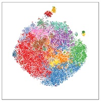
Brutally tough competition this year - the quality of science is just constantly rising year after year. I was really tempted by Orian's UMAP analysis (it looks like an elephant!):
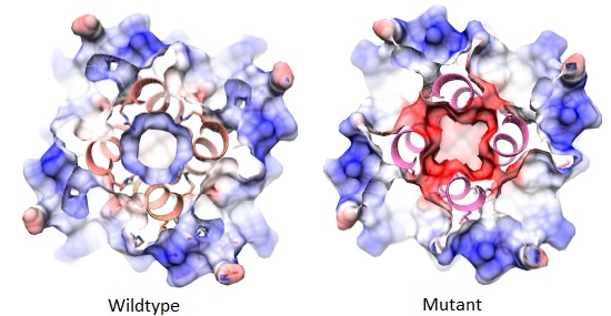
But for that single piece of data that just speaks for itself, it is hard to go past this crystal structure of a point mutation found in a novel primary immunodeficiency gene:
Well done Julika!

Julika receiving the Golden Pipette from past winner Lidia, in our virtual happy hour
Our lab retreat is a real highlight of the lab, with the Leuven and Cambridge teams getting together to share ideas and progress, brainstorm and socialise. We usually have a couple of retreats a year. Unfortunately our first was cancelled in 2020 by the lockdown, and we decided to make the second one virtual.
Having spent many, many hours on zoom calls over the past six months, I was quite worried about three solid days of a virtual zoom meeting. Fortunately it was a great success! Excellent science (of course) and the level of interaction was as good as an in-person retreat. I don't think it can ever replace in-person meetings, since the success builds on the already-existing personal connections, but perhaps a virtual retreat will substitute for every second retreat going forward?
Secret tips for a zoom retreat:


Just starting her PhD, Julika already has several major successes under her belt, including identifying a new primary immunodeficiency (stay tuned!) and spear-heading an open science study on COVID pathology.
We anticipate more great successes from Julika during her FWO fellowship!
Suikerrijke voeding veroorzaakt de ontwikkeling van alvleesklierkanker en verhoogt de kans op een dodelijke afloop. Tot die bevinding zijn onderzoekers van het Vlaams Instituut voor Biotechnologie (VIB) en de KU Leuven gekomen.
Een suikerrijk dieet verhoogt niet alleen de kans op het ontwikkelen van pancreaskanker. Het stimuleert ook de agressieve groei van tumoren. Tot die conclusie komen onderzoekers van VIB-KU Leuven en collega's van het Babraham Institute van de Britse universiteit Cambridge.
De resultaten zijn gepubliceerd in het tijdschrift Cell Reports.
Alvleesklierkanker is een zeldzame maar dodelijke vorm van kanker door late detectie en een slecht begrip van risicofactoren. Bekende risicofactoren zijn obesitas, voeding en diabetes type 2, maar door de lage incidentie en de onderlinge samenhang is hun individuele bijdrage moeilijk in te schatten.
De onderzoekers voltooiden een uitgebreid project met experimenteel werk bij muizen en menselijke gegevens. Bij de dieren werd de progressie van pancreaskanker beïnvloed door voedingssuiker, met een snellere tumorgroei en verhoogde dodelijkheid.
Het effect bij mensen blijkt hetzelfde. Bij 500 deelnemers aan de studie onderzochten de onderzoekers de interactie tussen genen en voeding en ontdekten dat hoge niveaus van voedingssuiker het risico op alvleesklierkanker verhoogden. 'We hebben jarenlang gekeken naar verschillende voedings- en genetische veranderingen en niets komt in de buurt van de nadelige gevolgen van een dieet met veel suiker', zegt James Dooley, senior wetenschapper bij het Babraham Institute.
Plantaardige voeding met een equivalent van een avocado per dag vermindert het risico op pancreaskanker met tien procent.
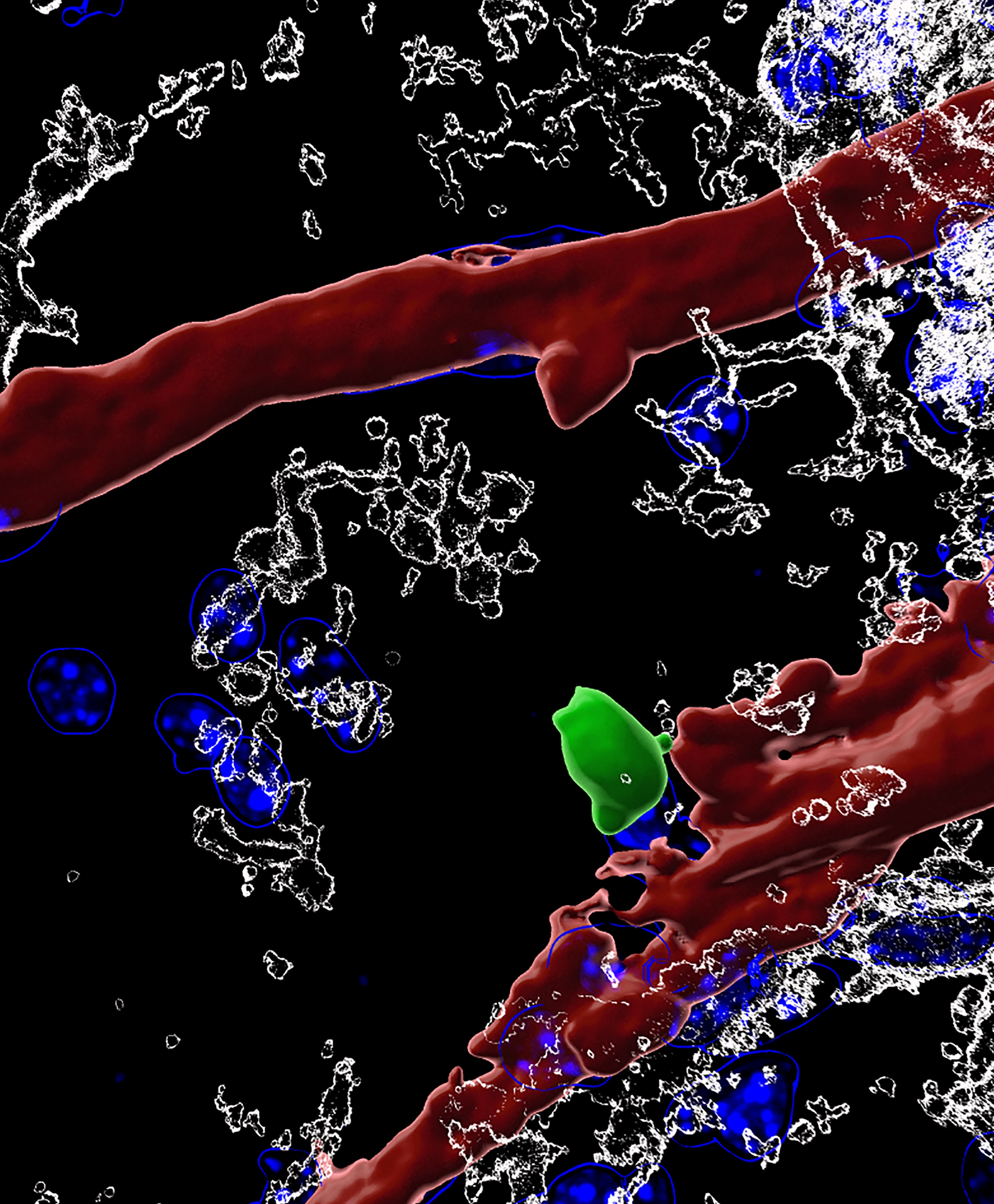
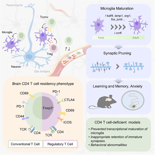
Whether white blood cells can be found in the brain has been controversial, and what they might be doing used to be complete mystery. In a seminal study published in Cell, an international team of scientists led by Prof. Adrian Liston (VIB-KU Leuven, Belgium & Babraham Institute, UK) describe a population of specialized brain-resident immune cells discovered in the mouse and human brain, and show that the presence of white blood cells is essential for normal brain development in mice.
 Like a highly fortified headquarters, our brain enjoys special protection from what is circulating in the rest of our body through the blood-brain barrier. This highly selective border makes sure that passage from the blood to the brain is tightly regulated.
The blood-brain barrier also separates the brain from our body’s immune system, which is why it has its own resident immune cells, called microglia, which trigger inflammation and tissue repair. Microglia arrive in the brain during embryonic development, and later on, the population becomes self-renewing.
Yet, white blood cells—which are part of our immune system—have been found to play a role in different brain diseases, including multiple sclerosis, Alzheimer’s and Parkinson’s disease or stroke. Whether or not white blood cells can be found in healthy brains as well, and what they might be doing there, has been subject of intense debate. An interdisciplinary team of scientists led by Prof. Adrian Liston (VIB-KU Leuven, Babraham Institute) set out to find the answers.
White blood cells in the brain
"A misconception about white blood cells comes from their name,” explains Dr. Oliver Burton (Babraham Institute). “These 'immune cells' are not just present in the blood. They are constantly circulating around our body and enter all of our organs, including—as it turns out—the brain. We are only just starting to discover what white blood cells do when they leave the blood. This research indicates that they act as a go-between, transferring information from the rest of the body to the brain environment"
The team quantified and characterized a small but distinct population of brain-resident T helper cells present in mouse and human brain tissue. T cells are a specific type of white blood cells specialized for scanning cell surfaces for evidence of infection and triggering an appropriate immune response. New technologies allowed the researchers to study the cells in great detail, including the processes by which circulating T cells entered the brain and began to develop the features of brain-resident T cells.
Dr. Carlos Roca (Babraham Institute): “Science is becoming increasingly multidisciplinary. Here, we didn't just bring in expertise from immunology, neuroscience and microbiology, but also from computer science and applied mathematics. New approaches for data analysis allow us to reach a much deeper level of understanding of the biology of the white blood cells we found in the brain.”
An evolutionary role
When T helper cells are absent from the brain, the scientists found that the resident immune cells – microglia – in the mouse brain remained suspended between a fetal and adult developmental state. Observationally, mice lacking brain T cells showed multiple changes in their behavior. The analysis points to an important role for brain-resident T cells in brain development. If T cells participate in normal brain development in mice, could the same be true in humans?
“In mice, the wave of entry of immune cells at birth triggers a switch in brain development,” says Liston. “Humans have a much longer gestation than mice though, and we don't know about the timing of immune cell entry into the brain. Does this occur before birth? Is it delayed until after birth? Did a change in timing of entry contribute to the evolution of enhanced cognitive capacity in humans?”
The findings open up a whole new range of questions about how the brain and our immune system interact. "It has been really exciting to work on this project. We are learning so much about how our immune system can alter our brain, and how our brain modifies our immune system. The two are far more interconnected than we previously thought," says Dr. Emanuela Pasciuto (VIB-KU Leuven).
The study also brings in a connection with the gut microbiome, says Liston: “There are now multiple links between the bacteria in our gut and different neurological conditions, but without any convincing explanations for what connects them. We show that white blood cells are modified by gut bacteria, and then take that information with them into the brain. This could be the route by which our gut microbiome influences the brain.”
Taken together, the results contribute towards the increasing recognition of the role of immune cells in the brain and shed new light on its involvement in a range of neurological diseases.
Check out the full article here
Witte bloedcellen maken deel uit van ons immuunsysteem dat ons tegen ziektes beschermt. Of ze ook in de hersenen terug te vinden zijn, en wat ze daar dan zouden doen bleef tot nog toe een raadsel. Een internationaal team van wetenschappers onder leiding van professor Adrian Liston (VIB-KU Leuven, België en Babraham Institute, VK) toont nu aan dat witte bloedcellen wel degelijk voorkomen in de hersenen van zowel muizen als mensen, en dat hun aanwezigheid essentieel is voor normale hersenontwikkeling. De resultaten verschijnen deze week in het prestigieuze vakblad Cell.
 Onze hersenen worden als een versterkte burcht ommuurd door de bloed-hersenbarrière. Die moet vermijden dat stoffen die in onze bloedbaan circuleren zomaar in onze hersenen terechtkomen. Via de bloed-hersenbarrière worden onder strikte controle enkel welbepaalde stoffen uitgewisseld.
De bloed-hersenbarrière schermt de hersenen ook af van ons immuunsysteem, dat de rest van ons lichaam patrouilleert om bijvoorbeeld bacteriële of virale indringers op te sporen en uit te schakelen. Precies daarom heeft het brein z’n eigen immuuncellen: microglia.
Toch blijken witte bloedcellen ook een rol te spelen bij verschillende hersenaandoeningen. Denk maar aan MS, alzheimer, parkinson of een beroerte. Hierbij gaat het wel telkens om ziek of ‘beschadigd’ hersenweefsel, waar mogelijk ook de bloed-hersenbarrière is aangetast. De vraag bleef dus of – en waarom – witte bloedcellen nu werkelijk aanwezig zijn in hersenen die normaal en gezond zijn.
Witte bloedcellen in de hersenen
Een interdisciplinair team van wetenschappers onder leiding van prof. Adrian Liston (VIB-KU Leuven, Babraham Institute) heeft nu een kleine maar belangrijke groep van T-helpercellen ontdekt in hersenweefsel afkomstig van muizen en van mensen. T-helpercellen zijn een specifiek type witte bloedcellen, gespecialiseerd in het scannen van celoppervlakken op aanwijzingen van infectie en in het op gang trekken van een aangepaste immuunreactie. Aan de hand van de laatste technologie konden de wetenschappers de T-helpercellen tot in detail bestuderen, inclusief hoe en wanneer ze in de hersenen terecht komen.
Dr. Emanuela Pasciuto (VIB-KU Leuven), postdoctoraal onderzoeker in het team van Liston benadrukt het belang van interdisciplinair onderzoek: “Om de rol van witte bloedcellen in het brein in kaart te brengen hebben we niet alleen expertise van immunologie, neurowetenschappen en microbiologie bij elkaar gebracht, maar ook van informatica en toegepaste wiskunde. Nieuwe benaderingen voor data-analyse stellen ons in staat om een veel dieper begrip te krijgen van de biologie van de witte bloedcellen die we in de hersenen hebben gevonden.”
 Een evolutionaire rol
De onderzoekers stelden vast dat in muizenhersenen zonder T-helpercellen de ontwikkeling van de typische immuuncellen van het brein (de microglia) bleef hangen ergens tussen een foetale en volwassen ontwikkelingsstatus. De muizen zonder T-helpercellen in de hersenen vertoonden bovendien verschillende gedragsafwijkingen, wat wijst op een belangrijke rol voor de T-helpercellen bij de normale hersenontwikkeling. En als dat geldt voor muizen, zou hetzelfde dan ook waar zijn voor mensen?
“We zien dat de toestroom van immuuncellen in de hersenen bij de geboorte van muizen leidt tot een omslag in het ontwikkelingsproces,” zegt Liston. “Maar de zwangerschap bij mensen is veel langer dan bij muizen, en we weten niet wanneer de immuuncellen dan toekomen in het menselijk brein. Gebeurt het nog vóór de geboorte? Is het uitgesteld tot na de geboorte? Kan een verandering in de timing bijgedragen hebben aan de evolutie van de uitzonderlijke hersencapaciteit van mensen?”
De bevindingen openen een heel nieuw gamma aan vragen over de wisselwerking tussen ons brein en ons immuunsysteem. “We leren nog elke dag bij over hoe ons immuunsysteem ons brein kan beïnvloeden en vice versa. De twee zijn veel meer met elkaar verbonden dan we eerder dachten,” zegt Pasciuto.
Darmen en hersenen
De studie legt ook nieuwe verbanden tussen ons brein en onze darmflora, aldus Liston: “Heel wat neurologische aandoeningen worden in verband gebracht met bacteriën in onze darmen, maar zonder overtuigende verklaringen voor die connectie. Onze resultaten laten zien dat darmbacteriën witte bloedcellen kunnen beïnvloeden, die deze ‘informatie’ vervolgens mee nemen naar de hersenen. Dit zou de manier kunnen zijn waarop onze darmflora onze hersenen beïnvloeden. ”
De nieuwe resultaten dragen enorm bij tot de groeiende kennis over de rol van immuuncellen in de hersenen, zowel tijdens de normale ontwikkeling als bij verschillende ziekteprocessen.
Research using mice and human data shows how diet affects disease risk
Eating a diet high in sugar increases the likelihood of developing pancreatic cancer in some individuals and also drives the aggressive growth of tumours, a study by researchers from the Babraham Institute, Cambridge, UK, and VIB-KU Leuven, Belgium, has found.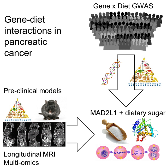
Pancreatic cancer is a rare but fatal form of cancer, due to late detection and a poor understanding of the risk factors. Known risk factors include obesity, diet and type 2 diabetes, however the low incidence rate and interconnection of these factors mean that it is difficult to tease apart their individual contribution.
The researchers first studied the effects of obesity, diet and diabetes on pancreatic cancer development, growth and lethality in mice. In parallel they analysed the effect of diet using data from the European Prospective Investigation into Cancer and Nutrition (EPIC) study, which followed over half a million Europeans for 20 years.
The researchers found that obesity, diet and diabetes had profound and differing impacts on cancer incidence and growth. Using mice, the results indicated that obesity, dietary animal fats and dietary sugar were independent drivers of different facets of pancreatic cancer progression. In particular the results shed light on how pancreatic cancer might be affected by dietary sugar, with more rapid tumour growth and escalated lethality.

The effect of dietary sugars on pancreatic cancer development was preserved between mice and humans. In 500 study participants with pancreatic cancer, the researchers explored the interaction between genes and diet and found that high levels of dietary sugar increased pancreatic cancer risk in individuals with a certain genetic variation (found in 6% of the population). Dr James DooleyDr James Dooley, senior staff scientist in the Immunology programme at the Babraham Institute, said: "Our study raises concern about the remarkable toxicity of sugar in our diet. We spent years looking at different dietary and genetic changes, and nothing has anywhere near the detrimental impact of a high sugar diet. Our findings suggest that it drives pancreatic cancer onset and makes it a more aggressive and lethal tumour."
Analysis of the human-derived data from the large EPIC study suggested that dietary plant fats reduced the risk of pancreatic cancer, estimating a 10% decrease in risk when eating the equivalent of an avocado a day.
Professor Adrian Liston, senior group leader at the Babraham Institute, said: "This study shows the power of combining animal research with the study of patients. We were able to use epidemiology and large patient-based resources to find a link in humans, and then go back to an animal model to formally test the direction of causality. Finding the same gene-diet link in both mice and humans makes us confident that diet is modifying disease risk, and gives us the tools to test preventative and therapeutic interventions."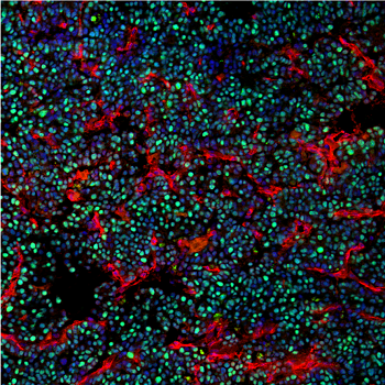 Proliferating pancreatic cancer cells in mice fed high sugar dietsWhile the impact of nutrition on cancer development continues to be explored and debated, this study provides a vital foundation for further studies and important indications to explore. Armed with this knowledge, clinicians could be supported to identify individuals at increased risk for screening and individuals with a high-risk genetic background could take pro-active dietary changes to reduce their risk of pancreatic cancer.
Ali Stunt FRSA, Founder and Chief Executive of Pancreatic Cancer Action, concurs: "Here at Pancreatic Cancer Action, we strongly advocate for high quality research into this neglected cancer. Dr Liston and his team have made a major contribution into understanding how diet and genetics changes the risk for pancreatic cancer. Healthy lifestyle choices, such as avoiding sugar-sweetened beverages and choosing a diet rich in vegetables, can reduce your risk of pancreatic cancer, and may be especially important in families with a history of the disease."